
CLASS 12 BIOLOGY · CHAPTER 2 · READ
Human Reproductionमानव जननHuman Reproduction
Male and female reproductive systems → gametogenesis → menstrual cycle → fertilisation and implantation → pregnancy → parturition and lactation. NCERT order, simple words.नर और मादा जनन तंत्र → युग्मकजनन → आर्तव चक्र → निषेचन और अंतर्रोपण → गर्भावस्था → प्रसव और दुग्धस्रवण। NCERT के क्रम में, आसान भाषा में।Male aur female reproductive system → gametogenesis → menstrual cycle → fertilisation aur implantation → pregnancy → parturition aur lactation. NCERT order, simple words.
Didn’t understand a paragraph? Tap it (or its अ button) → हिंदी / Hinglish / Simple English.कोई पैराग्राफ़ समझ नहीं आया? उस पर (या अ बटन पर) टैप करें → English / Hinglish / Simple English।Koi paragraph samajh nahi aaya? Us par (ya अ button par) tap karo → हिंदी / English / Simple English.
Diagrams in this chapterइस अध्याय के डायग्राम
 Seminiferous tubule (sectional view)शुक्रजनक नलिका (काट)Fig 2.2, 2.5
 Fertilisation, cleavage and implantationनिषेचन, विदलन और अंतर्रोपणFig 2.11
 Gametogenesis: spermatogenesis vs oogenesisयुग्मकजनन: शुक्राणुजनन व अंडजननFig 2.8
 Structure of a spermशुक्राणु की संरचनाFig 2.6
 Section of ovary: follicles to corpus luteumअंडाशय की काट: पुटिका से पीत पिंड तकFig 2.7
 Menstrual cycle (28 days)आर्तव चक्र (28 दिन)Fig 2.9 (schematic)
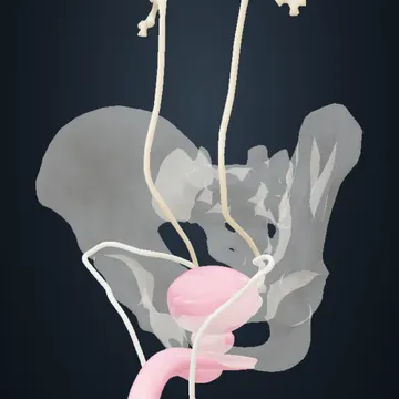 Human male reproductive system3D model ↗
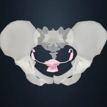 Human female reproductive system3D model ↗
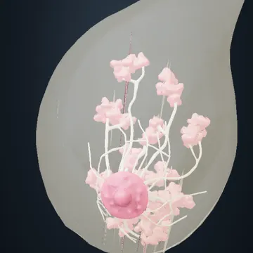 Human mammary gland3D model ↗
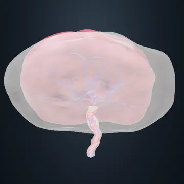 Human placenta and umbilical cord (full term)3D model ↗
In this chapter (NCERT sections)इस अध्याय में (NCERT खंड)
- 2.1The Male Reproductive Systemनर जनन तंत्रMale Reproductive System
- 2.2The Female Reproductive Systemमादा जनन तंत्रFemale Reproductive System
- 2.3Gametogenesisयुग्मकजननGametogenesis
- 2.4Menstrual Cycleआर्तव चक्रMenstrual Cycle
- 2.5Fertilisation and Implantationनिषेचन एवं अंतर्रोपणFertilisation aur Implantation
- 2.6Pregnancy and Embryonic Developmentसगर्भता एवं भ्रूणीय परिवर्धनPregnancy aur Embryonic Development
- 2.7Parturition and Lactationप्रसव एवं दुग्धस्रवणParturition aur Lactation
Humans reproduce sexually and are viviparous. The reproductive events are: making gametes (gametogenesis) — sperms in males and ovum in females; transfer of sperms into the female genital tract (insemination); fusion of the gametes (fertilisation) to form a zygote; formation of the blastocyst and its attachment to the uterine wall (implantation); development of the embryo (gestation); and delivery of the baby (parturition). These events begin after puberty. Sperm formation continues even in old men, but formation of ova stops in women around the age of fifty.मनुष्य लैंगिक जनन करते हैं और सजीवप्रजक हैं। जनन की घटनाएँ हैं: युग्मक बनना (युग्मकजनन) — पुरुष में शुक्राणु और स्त्री में अंडाणु; शुक्राणुओं का मादा जनन पथ में पहुँचना (वीर्यसेचन); युग्मकों का मिलकर युग्मनज बनाना (निषेचन); कोरकपुटी बनना और गर्भाशय की दीवार से जुड़ना (अंतर्रोपण); भ्रूण का विकास (सगर्भता); और शिशु का जन्म (प्रसव)। ये घटनाएँ यौवनारंभ के बाद शुरू होती हैं। शुक्राणु बनना बूढ़े पुरुषों में भी चलता रहता है, पर स्त्रियों में अंडाणु बनना लगभग पचास वर्ष की आयु में रुक जाता है।Humans sexually reproduce karte hain aur viviparous hain. Reproduction ke events hain: gametes banna (gametogenesis) — male mein sperms aur female mein ovum; sperms ka female genital tract mein pahunchna (insemination); gametes ka fuse hokar zygote banana (fertilisation); blastocyst banna aur uterus ki wall se judna (implantation); embryo ka development (gestation); aur baby ki delivery (parturition). Ye sab puberty ke baad shuru hota hai. Sperm banna boodhe aadmiyon mein bhi chalta rehta hai, par aurton mein ovum banna lagbhag 50 saal ki age mein ruk jaata hai.Humans make babies by sexual reproduction and give birth to live young. The steps are: make sperm and egg → sperm enters the female body → sperm and egg join → the early embryo sticks to the uterus → the baby grows → the baby is born.
2.1The Male Reproductive Systemनर जनन तंत्रMale Reproductive System
The male reproductive system lies in the pelvis region NCERT Fig 2.1a. It has a pair of testes3D, accessory ducts, accessory glands and the external genitalia — see the whole male reproductive system in 3D3D.नर जनन तंत्र श्रोणि (pelvis) क्षेत्र में होता है NCERT Fig 2.1a। इसमें एक जोड़ी वृषण3D, सहायक नलिकाएँ, सहायक ग्रंथियाँ और बाह्य जननांग होते हैं — पूरा नर जनन तंत्र 3D में3D देखें।Male reproductive system pelvis region mein hota hai NCERT Fig 2.1a. Isme ek jodi testes3D, accessory ducts, accessory glands aur external genitalia hote hain — poora male reproductive system 3D mein3D dekho.The male system sits in the lower belly (pelvis). It has two testes, tubes (ducts), glands and the penis.
Seminiferous tubule (sectional view)शुक्रजनक नलिका (काट)Seminiferous tubule (sectional view)NCERT Fig 2.2, 2.5 · 11 tappable partsOpenThe testes lie outside the abdominal cavity in a pouch called the scrotum, which keeps them 2–2.5 °C cooler than body temperature — needed for sperm formation. An adult testis is oval, about 4–5 cm long and 2–3 cm wide, with a dense covering. Each testis has about 250 testicular lobules, and each lobule has 1 to 3 highly coiled seminiferous tubules, where sperms are made NCERT Fig 2.1b. Each tubule is lined inside by male germ cells (spermatogonia) and Sertoli cells: germ cells divide by meiosis to form sperms, and Sertoli cells nourish the germ cells NCERT Fig 2.2. Outside the tubules, the interstitial spaces have small blood vessels and Leydig (interstitial) cells, which make and secrete androgens. Other immunologically competent cells are also present.वृषण पेट की गुहा के बाहर एक थैली — वृषणकोष — में रहते हैं, जो उन्हें शरीर के तापमान से 2–2.5 °C ठंडा रखती है — शुक्राणु बनने के लिए यह ज़रूरी है। वयस्क वृषण अंडाकार, लगभग 4–5 cm लंबा और 2–3 cm चौड़ा होता है, जिस पर घना आवरण होता है। हर वृषण में लगभग 250 वृषण पालिकाएँ होती हैं, और हर पालिका में 1 से 3 बहुत कुंडलित शुक्रजनक नलिकाएँ होती हैं, जिनमें शुक्राणु बनते हैं NCERT Fig 2.1b। हर नलिका अंदर से नर जनन कोशिकाओं (शुक्राणुजन) और सर्टोली कोशिकाओं से अस्तरित होती है: जनन कोशिकाएँ अर्धसूत्री विभाजन से शुक्राणु बनाती हैं, और सर्टोली कोशिकाएँ उन्हें पोषण देती हैं NCERT Fig 2.2। नलिकाओं के बाहर अंतराली स्थानों में छोटी रक्त वाहिकाएँ और लीडिग (अंतराली) कोशिकाएँ होती हैं, जो पुंजन (एंड्रोजन) बनाकर स्रावित करती हैं। यहाँ कुछ प्रतिरक्षा-सक्षम कोशिकाएँ भी होती हैं।Testes pet ki cavity ke bahar ek thaili — scrotum — mein rehte hain, jo unhe body temperature se 2–2.5 °C thanda rakhti hai — sperm banne ke liye ye zaroori hai. Adult testis oval, lagbhag 4–5 cm lamba aur 2–3 cm chauda hota hai, upar dense covering hoti hai. Har testis mein lagbhag 250 testicular lobules hote hain, aur har lobule mein 1 se 3 bahut coiled seminiferous tubules hote hain, jahan sperms bante hain NCERT Fig 2.1b. Har tubule andar se male germ cells (spermatogonia) aur Sertoli cells se lined hota hai: germ cells meiosis se sperms banati hain, aur Sertoli cells unhe nutrition deti hain NCERT Fig 2.2. Tubules ke bahar interstitial spaces mein chhoti blood vessels aur Leydig (interstitial) cells hoti hain, jo androgens banakar secrete karti hain. Kuch immunologically competent cells bhi hoti hain.The testes hang outside the body in the scrotum, which keeps them a little cooler — sperm need this. Each testis has about 250 small parts (lobules); each has 1–3 coiled tubes where sperm are made. Sertoli cells feed the young sperm cells. Leydig cells, between the tubes, make male hormones (androgens).
The male sex accessory ducts are the rete testis, vasa efferentia, epididymis3D and vas deferens3D NCERT Fig 2.1b. Seminiferous tubules open into the vasa efferentia through the rete testis. The vasa efferentia leave the testis and open into the epididymis, which lies along the back (posterior) surface of each testis. The epididymis leads to the vas deferens, which goes up into the abdomen and loops over the urinary bladder. It receives a duct from the seminal vesicle3D and opens into the urethra3D as the ejaculatory duct. These ducts store and carry sperms from the testis to the outside. The urethra starts from the urinary bladder and runs through the penis to its outer opening, the urethral meatus.नर सहायक नलिकाएँ हैं — वृषण जालिका (rete testis), शुक्रवाहिकाएँ (vasa efferentia), अधिवृषण (epididymis)3D और शुक्रवाहक (vas deferens)3D NCERT Fig 2.1b। शुक्रजनक नलिकाएँ वृषण जालिका के रास्ते शुक्रवाहिकाओं में खुलती हैं। शुक्रवाहिकाएँ वृषण से निकलकर अधिवृषण में खुलती हैं, जो हर वृषण की पिछली सतह के साथ होता है। अधिवृषण आगे शुक्रवाहक बनता है, जो ऊपर पेट में जाकर मूत्राशय के ऊपर से घूमता है। इसमें शुक्राशय3D की एक नलिका आकर मिलती है और यह स्खलनीय वाहिनी (ejaculatory duct) बनकर मूत्रमार्ग3D में खुलता है। ये नलिकाएँ शुक्राणुओं को जमा करती हैं और वृषण से बाहर तक ले जाती हैं। मूत्रमार्ग मूत्राशय से शुरू होकर शिश्न से होते हुए उसके बाहरी छिद्र — मूत्रमार्ग मुख (urethral meatus) — तक जाता है।Male accessory ducts hain — rete testis, vasa efferentia, epididymis3D aur vas deferens3D NCERT Fig 2.1b. Seminiferous tubules rete testis ke through vasa efferentia mein khulte hain. Vasa efferentia testis se nikalkar epididymis mein khulti hain, jo har testis ki peeche wali surface ke saath hota hai. Epididymis aage vas deferens banta hai, jo upar abdomen mein jaakar urinary bladder ke upar loop banata hai. Isme seminal vesicle3D ki duct aakar milti hai aur ye ejaculatory duct banke urethra3D mein khulta hai. Ye ducts sperms ko store karti hain aur testis se bahar tak le jaati hain. Urethra urinary bladder se shuru hokar penis se hote hue bahar wale opening — urethral meatus — tak jaati hai.Sperm travel through a chain of tubes: rete testis → vasa efferentia → epididymis → vas deferens → ejaculatory duct → urethra → outside. These tubes store and carry the sperm.
The penis3D is the male external genitalia NCERT Fig 2.1a, b. It is made of special tissue that helps in its erection to make insemination possible. Its enlarged end, the glans penis, is covered by a loose fold of skin called the foreskin. The male accessory glands are the paired seminal vesicles3D, a prostate3D and the paired bulbourethral glands. Their secretions form the seminal plasma, rich in fructose, calcium and certain enzymes. The bulbourethral secretions also help in lubrication of the penis.शिश्न3D नर का बाह्य जननांग है NCERT Fig 2.1a, b। यह विशेष ऊतक से बना होता है जो वीर्यसेचन के लिए उसके उत्थान में मदद करता है। इसका फूला हुआ सिरा — शिश्नमुंड (glans penis) — त्वचा की ढीली परत अग्रच्छद (foreskin) से ढका होता है। नर सहायक ग्रंथियाँ हैं — एक जोड़ी शुक्राशय3D, एक पुरःस्थ ग्रंथि (prostate)3D और एक जोड़ी बल्बोयूरेथ्रल ग्रंथियाँ। इनके स्राव से शुक्रीय प्लाज़्मा बनता है, जिसमें फ्रक्टोज़, कैल्शियम और कुछ एंजाइम भरपूर होते हैं। बल्बोयूरेथ्रल ग्रंथियों का स्राव शिश्न को चिकना भी करता है।Penis3D male ka external genitalia hai NCERT Fig 2.1a, b. Ye special tissue se bana hota hai jo insemination ke liye erection mein help karta hai. Iska phoola hua end — glans penis — skin ki dheeli layer foreskin se dhaka hota hai. Male accessory glands hain — ek jodi seminal vesicles3D, ek prostate3D aur ek jodi bulbourethral glands. Inke secretion se seminal plasma banta hai, jisme fructose, calcium aur kuch enzymes bhare hote hain. Bulbourethral glands ka secretion penis ko lubricate bhi karta hai.The penis is the outer male organ. Three kinds of glands (seminal vesicles, prostate, bulbourethral glands) make a fluid called seminal plasma. It has sugar (fructose), calcium and enzymes for the sperm.
2.2The Female Reproductive Systemमादा जनन तंत्रFemale Reproductive System
The female reproductive system has a pair of ovaries3D, a pair of oviducts, the uterus, cervix, vagina and the external genitalia, in the pelvic region NCERT Fig 2.3a. Together with a pair of mammary glands3D, these parts work together to support ovulation, fertilisation, pregnancy, birth and child care — see the female reproductive system in 3D3D.मादा जनन तंत्र में एक जोड़ी अंडाशय3D, एक जोड़ी अंडवाहिनियाँ, गर्भाशय, ग्रीवा (cervix), योनि और बाह्य जननांग होते हैं, जो श्रोणि क्षेत्र में हैं NCERT Fig 2.3a। एक जोड़ी स्तन ग्रंथियों3D के साथ ये सब मिलकर अंडोत्सर्ग, निषेचन, गर्भावस्था, जन्म और शिशु की देखभाल में मदद करते हैं — मादा जनन तंत्र 3D में3D देखें।Female reproductive system mein ek jodi ovaries3D, ek jodi oviducts, uterus, cervix, vagina aur external genitalia hote hain, pelvic region mein NCERT Fig 2.3a. Ek jodi mammary glands3D ke saath ye sab milkar ovulation, fertilisation, pregnancy, birth aur child care mein help karte hain — female reproductive system 3D mein3D dekho.The female system has two ovaries, two tubes (oviducts), the uterus, cervix, vagina and outer parts. With the breasts (mammary glands), they help in egg release, fertilisation, pregnancy, birth and baby care.
Ovaries are the primary female sex organs: they make the female gamete (ovum) and several steroid hormones (ovarian hormones). One ovary lies on each side of the lower abdomen NCERT Fig 2.3b. Each is about 2–4 cm long and is joined to the pelvic wall and uterus by ligaments. It is covered by a thin epithelium that encloses the ovarian stroma, which has two zones — an outer cortex and an inner medulla.अंडाशय प्राथमिक मादा जनन अंग हैं: ये मादा युग्मक (अंडाणु) और कई स्टेरॉयड हॉर्मोन (अंडाशयी हॉर्मोन) बनाते हैं। पेट के निचले भाग में दोनों ओर एक-एक अंडाशय होता है NCERT Fig 2.3b। हर अंडाशय लगभग 2–4 cm लंबा होता है और स्नायुओं (ligaments) से श्रोणि की दीवार और गर्भाशय से जुड़ा रहता है। इस पर पतली उपकला होती है जो अंडाशयी पीठिका (stroma) को घेरती है; पीठिका के दो भाग हैं — बाहरी वल्कुट (cortex) और भीतरी मध्यांश (medulla)।Ovaries primary female sex organs hain: ye female gamete (ovum) aur kai steroid hormones (ovarian hormones) banati hain. Lower abdomen mein dono side ek-ek ovary hoti hai NCERT Fig 2.3b. Har ovary lagbhag 2–4 cm lambi hoti hai aur ligaments se pelvic wall aur uterus se judi rehti hai. Iske upar patla epithelium hota hai jo ovarian stroma ko gherta hai; stroma ke do zones hain — bahar cortex aur andar medulla.The ovaries make eggs and female hormones. There is one on each side, about 2–4 cm long. Inside, the ovary has an outer part (cortex) and an inner part (medulla).
The oviducts (fallopian tubes), uterus and vagina are the female accessory ducts. Each fallopian tube is about 10–12 cm long and runs from near the ovary to the uterus NCERT Fig 2.3b. The part closest to the ovary is the funnel-shaped infundibulum, whose edges have finger-like fimbriae that collect the ovum after ovulation. It leads to a wider part, the ampulla, and the last narrow part, the isthmus, joins the uterus.अंडवाहिनियाँ (फैलोपियन नलिकाएँ), गर्भाशय और योनि मादा सहायक नलिकाएँ हैं। हर फैलोपियन नलिका लगभग 10–12 cm लंबी होती है और अंडाशय के पास से गर्भाशय तक जाती है NCERT Fig 2.3b। अंडाशय के सबसे पास वाला कीप जैसा भाग कीपक (infundibulum) है, जिसके किनारों पर उँगली जैसी झालर (fimbriae) होती हैं जो अंडोत्सर्ग के बाद अंडाणु को पकड़ती हैं। आगे चौड़ा भाग तुंबिका (ampulla) है, और अंतिम पतला भाग संकीर्ण पथ (isthmus) गर्भाशय से जुड़ता है।Oviducts (fallopian tubes), uterus aur vagina female accessory ducts hain. Har fallopian tube lagbhag 10–12 cm lambi hoti hai aur ovary ke paas se uterus tak jaati hai NCERT Fig 2.3b. Ovary ke sabse paas wala funnel jaisa part infundibulum hai, jiske edges par ungli jaisi fimbriae hoti hain jo ovulation ke baad ovum ko pakadti hain. Aage chauda part ampulla hai, aur last patla part isthmus uterus se judta hai.Each fallopian tube is 10–12 cm long. Its funnel end (infundibulum) has finger-like fimbriae that catch the egg. Then comes the wide ampulla and the narrow isthmus, which joins the uterus.
The uterus3D (womb) is single and shaped like an inverted pear, held by ligaments attached to the pelvic wall. It opens into the vagina through a narrow cervix3D. The cavity of the cervix, the cervical canal, together with the vagina forms the birth canal. The uterine wall has three layers: an outer thin perimetrium, a middle thick layer of smooth muscle, the myometrium, and an inner glandular layer, the endometrium, which lines the uterine cavity. The endometrium changes in a cycle during the menstrual cycle, while the myometrium contracts strongly during delivery of the baby.गर्भाशय3D एक ही होता है, आकार उल्टी नाशपाती जैसा, और श्रोणि की दीवार से जुड़े स्नायुओं से टिका रहता है। यह संकरी ग्रीवा (cervix)3D के रास्ते योनि में खुलता है। ग्रीवा की गुहा — ग्रीवा नाल (cervical canal) — योनि के साथ मिलकर जन्म नाल (birth canal) बनाती है। गर्भाशय की दीवार में तीन परतें हैं: बाहरी पतली गर्भाशय परिस्तर (perimetrium), बीच की मोटी चिकनी पेशी की परत गर्भाशय पेशीस्तर (myometrium), और भीतरी ग्रंथिल परत गर्भाशय अंतःस्तर (endometrium), जो गर्भाशय की गुहा को अस्तरित करती है। आर्तव चक्र में अंतःस्तर में चक्रीय बदलाव होते हैं, जबकि शिशु के जन्म के समय पेशीस्तर ज़ोर से सिकुड़ता है।Uterus3D (womb) ek hi hota hai, shape ulti nashpati (inverted pear) jaisa, aur pelvic wall se jude ligaments se tika rehta hai. Ye patli cervix3D ke through vagina mein khulta hai. Cervix ki cavity — cervical canal — vagina ke saath milkar birth canal banati hai. Uterus ki wall mein teen layers hain: bahar patli perimetrium, beech mein moti smooth muscle layer myometrium, aur andar glandular layer endometrium, jo uterus ki cavity ko line karti hai. Menstrual cycle mein endometrium cyclic changes dikhata hai, aur baby ki delivery ke time myometrium zor se contract karta hai.The uterus is shaped like an upside-down pear. It opens into the vagina through the cervix; the cervix canal and vagina form the birth canal. Its wall has 3 layers: perimetrium (outer), myometrium (muscle, pushes the baby out at birth), endometrium (inner lining, changes every month).
The female external genitalia are the mons pubis, labia majora, labia minora, hymen and clitoris NCERT Fig 2.3a. The mons pubis is a cushion of fatty tissue covered by skin and pubic hair. The labia majora are fleshy folds that extend down from the mons pubis and surround the vaginal opening; the labia minora are paired folds under them. The vaginal opening is often partly covered by a membrane, the hymen. The clitoris is a tiny finger-like structure at the upper junction of the two labia minora, above the urethral opening. The hymen may tear during the first coitus, but it can also break by a sudden fall or jolt, use of a tampon, or sports like horse riding and cycling, and in some women it stays even after coitus. So the presence or absence of the hymen is not a reliable indicator of virginity or sexual experience.मादा बाह्य जननांग हैं — जघन शैल (mons pubis), वृहद् भगोष्ठ (labia majora), लघु भगोष्ठ (labia minora), योनिच्छद (hymen) और भगशेफ (clitoris) NCERT Fig 2.3a। जघन शैल त्वचा और जघन बालों से ढका वसा ऊतक का गद्दा है। वृहद् भगोष्ठ मांसल परतें हैं जो जघन शैल से नीचे फैलकर योनि द्वार को घेरती हैं; लघु भगोष्ठ इनके नीचे की जोड़ीदार परतें हैं। योनि का द्वार अक्सर एक झिल्ली — योनिच्छद — से आंशिक रूप से ढका रहता है। भगशेफ दोनों लघु भगोष्ठों के ऊपरी जोड़ पर, मूत्र द्वार के ऊपर, एक छोटी उँगली जैसी रचना है। योनिच्छद पहले मैथुन में फट सकता है, पर यह अचानक गिरने या झटके, टैम्पोन के उपयोग, या घुड़सवारी और साइकिल चलाने जैसे खेलों से भी टूट सकता है, और कुछ स्त्रियों में मैथुन के बाद भी बना रहता है। इसलिए योनिच्छद का होना या न होना कौमार्य या यौन अनुभव का विश्वसनीय संकेत नहीं है।Female external genitalia hain — mons pubis, labia majora, labia minora, hymen aur clitoris NCERT Fig 2.3a. Mons pubis skin aur pubic hair se dhaka fat tissue ka cushion hai. Labia majora fleshy folds hain jo mons pubis se neeche failkar vaginal opening ko gherti hain; labia minora inke neeche ki paired folds hain. Vaginal opening aksar ek membrane — hymen — se partly dhaka hota hai. Clitoris dono labia minora ke upar wale junction par, urethral opening ke upar, ek chhoti ungli jaisi structure hai. Hymen pehle coitus mein phat sakta hai, par achanak girne ya jhatke, tampon use karne, ya horse riding aur cycling jaise sports se bhi toot sakta hai, aur kuch aurton mein coitus ke baad bhi rehta hai. Isliye hymen hona ya na hona virginity ya sexual experience ka reliable indicator nahi hai.The outer female parts are the mons pubis, labia majora, labia minora, hymen and clitoris. The hymen can break in many ways (a fall, sports, a tampon) or stay intact, so it tells nothing reliable about a woman's sexual experience.
A functional mammary gland3D is a feature of all female mammals. The mammary glands (breasts) are paired and contain glandular tissue and a variable amount of fat. The glandular tissue of each breast is divided into 15–20 mammary lobes3D with clusters of cells called alveoli NCERT Fig 2.4. Alveolar cells secrete milk, which is stored in the lumens of the alveoli. The alveoli open into mammary tubules; the tubules of each lobe join to form a mammary duct3D. Several mammary ducts join to form a wider mammary ampulla, which connects to the lactiferous duct and opens at the nipple3D, from where milk comes out during breastfeeding.क्रियाशील स्तन ग्रंथि3D सभी मादा स्तनधारियों का लक्षण है। स्तन ग्रंथियाँ (स्तन) जोड़ी में होती हैं और इनमें ग्रंथिल ऊतक और अलग-अलग मात्रा में वसा होती है। हर स्तन का ग्रंथिल ऊतक 15–20 स्तन पालियों3D में बँटा होता है, जिनमें कोशिकाओं के गुच्छे — कूपिकाएँ (alveoli) — होते हैं NCERT Fig 2.4। कूपिकाओं की कोशिकाएँ दूध स्रावित करती हैं, जो कूपिकाओं की गुहा में जमा होता है। कूपिकाएँ स्तन नलिकाओं (mammary tubules) में खुलती हैं; हर पाली की नलिकाएँ मिलकर एक स्तन वाहिनी3D बनाती हैं। कई स्तन वाहिनियाँ मिलकर चौड़ी स्तन तुंबिका (mammary ampulla) बनाती हैं, जो दुग्ध वाहिनी (lactiferous duct) से जुड़कर चूचुक3D पर खुलती है, जहाँ से स्तनपान के समय दूध निकलता है।Functional mammary gland3D saare female mammals ki pehchaan hai. Mammary glands (breasts) jodi mein hoti hain aur inme glandular tissue aur alag-alag amount mein fat hota hai. Har breast ka glandular tissue 15–20 mammary lobes3D mein bata hota hai, jinme cells ke clusters — alveoli — hote hain NCERT Fig 2.4. Alveoli ki cells milk secrete karti hain, jo alveoli ke lumen mein store hota hai. Alveoli mammary tubules mein khulti hain; har lobe ki tubules milkar ek mammary duct3D banati hain. Kai mammary ducts milkar chaudi mammary ampulla banati hain, jo lactiferous duct se judkar nipple3D par khulti hai, jahan se breastfeeding ke time milk nikalta hai.Each breast has 15–20 lobes. Small sacs (alveoli) make milk. Milk flows: alveoli → tubules → mammary duct → mammary ampulla → lactiferous duct → nipple.
2.3Gametogenesisयुग्मकजननGametogenesis
Gametogenesis: spermatogenesis vs oogenesisयुग्मकजनन: शुक्राणुजनन व अंडजननGametogenesis: spermatogenesis vs oogenesisNCERT Fig 2.8 · 11 tappable parts · 5 stepsOpenThe primary sex organs — testes in males and ovaries in females — make gametes (sperms and ovum) by gametogenesis. In the testis, immature male germ cells (spermatogonia) make sperms by spermatogenesis, which begins at puberty. Spermatogonia on the inner wall of the seminiferous tubules multiply by mitosis; each is diploid with 46 chromosomes. Some of them, called primary spermatocytes, periodically undergo meiosis. A primary spermatocyte completes meiosis I (reduction division) to form two equal, haploid secondary spermatocytes with 23 chromosomes each. These undergo meiosis II to give four equal, haploid spermatids NCERT Fig 2.5. Spermatids change into sperms by spermiogenesis. After that, sperm heads get embedded in the Sertoli cells, and the sperms are finally released from the tubules by spermiation.प्राथमिक जनन अंग — पुरुष में वृषण और स्त्री में अंडाशय — युग्मकजनन द्वारा युग्मक (शुक्राणु और अंडाणु) बनाते हैं। वृषण में अपरिपक्व नर जनन कोशिकाएँ (शुक्राणुजन) शुक्राणुजनन से शुक्राणु बनाती हैं, जो यौवनारंभ पर शुरू होता है। शुक्रजनक नलिकाओं की भीतरी दीवार पर शुक्राणुजन समसूत्री विभाजन से संख्या बढ़ाते हैं; हर शुक्राणुजन द्विगुणित होता है, जिसमें 46 गुणसूत्र होते हैं। इनमें से कुछ — प्राथमिक शुक्राणुकोशिकाएँ — समय-समय पर अर्धसूत्री विभाजन करती हैं। प्राथमिक शुक्राणुकोशिका अर्धसूत्री-I (न्यूनीकरण विभाजन) पूरा करके दो बराबर, अगुणित द्वितीयक शुक्राणुकोशिकाएँ बनाती है, जिनमें 23-23 गुणसूत्र होते हैं। ये अर्धसूत्री-II से चार बराबर, अगुणित शुक्राणुप्रसू (spermatids) बनाती हैं NCERT Fig 2.5। शुक्राणुप्रसू शुक्राणुकायांतरण से शुक्राणु बनते हैं। इसके बाद शुक्राणुओं के सिर सर्टोली कोशिकाओं में धँसे रहते हैं, और अंत में शुक्राणुमोचन से नलिकाओं से बाहर निकलते हैं।Primary sex organs — male mein testes aur female mein ovaries — gametogenesis se gametes (sperms aur ovum) banate hain. Testis mein immature male germ cells (spermatogonia) spermatogenesis se sperms banati hain, jo puberty par shuru hota hai. Seminiferous tubules ki andar wali wall par spermatogonia mitosis se number badhate hain; har spermatogonium diploid hota hai, 46 chromosomes wala. Inme se kuch — primary spermatocytes — time-time par meiosis karti hain. Primary spermatocyte meiosis I (reduction division) poora karke do equal, haploid secondary spermatocytes banati hai, har ek mein 23 chromosomes. Ye meiosis II se chaar equal, haploid spermatids banati hain NCERT Fig 2.5. Spermatids spermiogenesis se sperms bante hain. Iske baad sperms ke heads Sertoli cells mein dhanse rehte hain, aur last mein spermiation se tubules se release hote hain.Sperm making starts at puberty. Spermatogonium (46 chromosomes) → primary spermatocyte → meiosis I → 2 secondary spermatocytes (23 each) → meiosis II → 4 spermatids → spermiogenesis → 4 sperms → released by spermiation.
Hormones control spermatogenesis. At puberty the hypothalamus increases secretion of GnRH (gonadotropin releasing hormone). GnRH acts on the anterior pituitary, which releases two gonadotropins — LH (luteinising hormone) and FSH (follicle stimulating hormone). LH acts on the Leydig cells and makes them produce androgens, and androgens stimulate spermatogenesis. FSH acts on the Sertoli cells and makes them secrete some factors that help in spermiogenesis.शुक्राणुजनन हॉर्मोन से नियंत्रित होता है। यौवनारंभ पर हाइपोथैलेमस GnRH (गोनैडोट्रॉपिन रिलीज़िंग हॉर्मोन) का स्राव बढ़ाता है। GnRH अग्र पीयूष ग्रंथि पर काम करता है, जो दो गोनैडोट्रॉपिन छोड़ती है — LH (ल्यूटिनाइज़िंग हॉर्मोन) और FSH (पुटिका प्रेरक हॉर्मोन)। LH लीडिग कोशिकाओं पर काम करके पुंजन बनवाता है, और पुंजन शुक्राणुजनन को प्रेरित करते हैं। FSH सर्टोली कोशिकाओं पर काम करके कुछ कारक स्रावित करवाता है जो शुक्राणुकायांतरण में मदद करते हैं।Spermatogenesis hormones se control hota hai. Puberty par hypothalamus GnRH (gonadotropin releasing hormone) ka secretion badhata hai. GnRH anterior pituitary par kaam karta hai, jo do gonadotropins chhodti hai — LH aur FSH. LH Leydig cells par kaam karke androgens banwaata hai, aur androgens spermatogenesis ko stimulate karte hain. FSH Sertoli cells par kaam karke kuch factors secrete karwaata hai jo spermiogenesis mein help karte hain.Brain hormone GnRH → pituitary releases LH and FSH. LH → Leydig cells → androgens → sperm making. FSH → Sertoli cells → help sperm mature.
Structure of a spermशुक्राणु की संरचनाStructure of a spermNCERT Fig 2.6 · 6 tappable partsOpenA sperm is microscopic and has a head, neck, middle piece and tail NCERT Fig 2.6. A plasma membrane covers the whole body. The head has an elongated haploid nucleus; its front part is covered by a cap-like acrosome, filled with enzymes that help in fertilising the ovum. The middle piece has many mitochondria, which give energy for the movement of the tail — sperm motility is essential for fertilisation. A man ejaculates about 200–300 million sperms during a coitus; for normal fertility at least 60 per cent must have normal shape and size and at least 40 per cent must show vigorous motility.शुक्राणु सूक्ष्म होता है और इसमें सिर, ग्रीवा, मध्य भाग और पूँछ होते हैं NCERT Fig 2.6। पूरा शरीर प्लाज़्मा झिल्ली से ढका होता है। सिर में लंबा अगुणित केंद्रक होता है; उसका आगे का भाग टोपी जैसे एक्रोसोम (अग्रपिंडक) से ढका होता है, जिसमें अंडाणु के निषेचन में मदद करने वाले एंजाइम भरे होते हैं। मध्य भाग में बहुत सारे माइटोकॉन्ड्रिया होते हैं, जो पूँछ की गति के लिए ऊर्जा देते हैं — निषेचन के लिए शुक्राणु का गतिशील होना ज़रूरी है। एक मैथुन में पुरुष लगभग 20–30 करोड़ (200–300 million) शुक्राणु स्खलित करता है; सामान्य जनन क्षमता के लिए कम से कम 60 प्रतिशत शुक्राणुओं का आकार-माप सामान्य और कम से कम 40 प्रतिशत में तेज़ गतिशीलता होनी चाहिए।Sperm microscopic hota hai aur isme head, neck, middle piece aur tail hote hain NCERT Fig 2.6. Poori body plasma membrane se dhaki hoti hai. Head mein lamba haploid nucleus hota hai; uska aage wala part topi jaise acrosome se dhaka hota hai, jisme ovum ke fertilisation mein help karne wale enzymes bhare hote hain. Middle piece mein bahut saare mitochondria hote hain, jo tail ki movement ke liye energy dete hain — fertilisation ke liye sperm motility zaroori hai. Ek coitus mein male lagbhag 200–300 million sperms ejaculate karta hai; normal fertility ke liye kam se kam 60% sperms ka shape-size normal aur kam se kam 40% mein vigorous motility honi chahiye.A sperm has a head (nucleus + acrosome with enzymes), neck, middle piece (mitochondria = energy) and tail (to swim). About 200–300 million sperms are released at a time; at least 60% should be normal in shape and 40% should swim strongly.
Sperms released from the seminiferous tubules are carried by the accessory ducts. Secretions of the epididymis, vas deferens, seminal vesicle and prostate are needed for maturation and motility of sperms. The seminal plasma along with the sperms makes the semen. The work of the male accessory ducts and glands is kept up by the testicular hormones (androgens).शुक्रजनक नलिकाओं से निकले शुक्राणु सहायक नलिकाओं द्वारा आगे ले जाए जाते हैं। अधिवृषण, शुक्रवाहक, शुक्राशय और पुरःस्थ ग्रंथि के स्राव शुक्राणुओं के परिपक्वन और गतिशीलता के लिए ज़रूरी हैं। शुक्रीय प्लाज़्मा और शुक्राणु मिलकर वीर्य बनाते हैं। नर सहायक नलिकाओं और ग्रंथियों का काम वृषण हॉर्मोन (पुंजन) से बना रहता है।Seminiferous tubules se nikle sperms accessory ducts dwara aage le jaaye jaate hain. Epididymis, vas deferens, seminal vesicle aur prostate ke secretions sperms ki maturation aur motility ke liye zaroori hain. Seminal plasma aur sperms milkar semen banate hain. Male accessory ducts aur glands ka kaam testicular hormones (androgens) se chalta rehta hai.The ducts and glands help sperm mature and swim. Sperms + seminal plasma = semen. Androgens keep these ducts and glands working.
Section of ovary: follicles to corpus luteumअंडाशय की काट: पुटिका से पीत पिंड तकSection of ovary: follicles to corpus luteumNCERT Fig 2.7 · 9 tappable parts · 6 stepsOpenOogenesis is the formation of a mature female gamete, and it is very different from spermatogenesis. It starts during embryonic development: a couple of million gamete mother cells (oogonia) form in each fetal ovary, and no more oogonia are formed after birth. These cells start dividing, enter prophase-I of meiosis and get temporarily arrested there — they are now primary oocytes. Each primary oocyte gets a layer of granulosa cells around it and is called a primary follicle NCERT Fig 2.7. Many follicles degenerate between birth and puberty, so at puberty only 60,000–80,000 primary follicles are left in each ovary.अंडजनन (Oogenesis) परिपक्व मादा युग्मक बनने की प्रक्रिया है, और यह शुक्राणुजनन से काफ़ी अलग है। यह भ्रूणीय विकास के समय शुरू होता है: हर भ्रूणीय अंडाशय में लगभग बीस लाख युग्मक मातृ कोशिकाएँ (अंडजनी, oogonia) बनती हैं, और जन्म के बाद कोई नई अंडजनी नहीं बनती। ये कोशिकाएँ विभाजन शुरू करके अर्धसूत्री विभाजन की पूर्वावस्था-I (prophase-I) में पहुँचती हैं और वहीं अस्थायी रूप से रुक जाती हैं — अब ये प्राथमिक अंडक हैं। हर प्राथमिक अंडक के चारों ओर कणिकामय (granulosa) कोशिकाओं की एक परत बनती है और उसे प्राथमिक पुटक कहते हैं NCERT Fig 2.7। जन्म से यौवनारंभ तक बहुत से पुटक नष्ट हो जाते हैं, इसलिए यौवनारंभ पर हर अंडाशय में केवल 60,000–80,000 प्राथमिक पुटक बचते हैं।Oogenesis mature female gamete banne ka process hai, aur ye spermatogenesis se kaafi alag hai. Ye embryonic development ke time shuru hota hai: har fetal ovary mein lagbhag 2 million gamete mother cells (oogonia) banti hain, aur birth ke baad koi nayi oogonia nahi banti. Ye cells divide hokar meiosis ke prophase-I mein pahunchti hain aur wahin temporarily arrest ho jaati hain — ab ye primary oocytes hain. Har primary oocyte ke chaaron taraf granulosa cells ki ek layer banti hai aur use primary follicle kehte hain NCERT Fig 2.7. Birth se puberty tak bahut saare follicles degenerate ho jaate hain, isliye puberty par har ovary mein sirf 60,000–80,000 primary follicles bachte hain.Egg making starts before birth. All oogonia are made in the fetal ovary; no new ones later. They stop at an early stage of meiosis (primary oocytes) inside primary follicles. By puberty only 60,000–80,000 follicles are left in each ovary.
Primary follicles get more layers of granulosa cells and a new theca — now secondary follicles. A secondary follicle soon becomes a tertiary follicle, which has a fluid-filled cavity, the antrum; its theca becomes an inner theca interna and an outer theca externa. At this stage the primary oocyte grows and completes meiosis I. This division is unequal: it gives a large haploid secondary oocyte and a tiny first polar body NCERT Fig 2.8b. The secondary oocyte keeps most of the nutrient-rich cytoplasm. The tertiary follicle then becomes the mature Graafian follicle, and the secondary oocyte forms a new membrane, the zona pellucida, around itself. The Graafian follicle ruptures and releases the secondary oocyte (ovum) from the ovary — ovulation.प्राथमिक पुटकों के चारों ओर कणिकामय कोशिकाओं की और परतें और एक नया प्रावरक (theca) बनता है — अब ये द्वितीयक पुटक हैं। द्वितीयक पुटक जल्दी ही तृतीयक पुटक बन जाता है, जिसमें द्रव से भरी गुहा — गह्वर (antrum) — होती है; इसका प्रावरक भीतरी अंतः प्रावरक (theca interna) और बाहरी बाह्य प्रावरक (theca externa) में बँट जाता है। इसी अवस्था में प्राथमिक अंडक बड़ा होकर अर्धसूत्री-I पूरा करता है। यह विभाजन असमान होता है: एक बड़ा अगुणित द्वितीयक अंडक और एक छोटा प्रथम ध्रुवीय पिंड बनता है NCERT Fig 2.8b। द्वितीयक अंडक पोषक तत्वों से भरा ज़्यादातर कोशिकाद्रव्य अपने पास रखता है। फिर तृतीयक पुटक परिपक्व ग्राफी पुटक बनता है, और द्वितीयक अंडक अपने चारों ओर नई झिल्ली — पारदर्शी अंडावरण (zona pellucida) — बनाता है। ग्राफी पुटक फटता है और द्वितीयक अंडक (अंडाणु) अंडाशय से बाहर निकलता है — अंडोत्सर्ग।Primary follicles ke chaaron taraf granulosa cells ki aur layers aur ek nayi theca banti hai — ab ye secondary follicles hain. Secondary follicle jaldi hi tertiary follicle ban jaata hai, jisme fluid wali cavity — antrum — hoti hai; iski theca andar theca interna aur bahar theca externa mein bant jaati hai. Isi stage par primary oocyte bada hokar meiosis I poora karta hai. Ye division unequal hota hai: ek bada haploid secondary oocyte aur ek chhota first polar body banta hai NCERT Fig 2.8b. Secondary oocyte nutrient wala zyaadatar cytoplasm apne paas rakhta hai. Phir tertiary follicle mature Graafian follicle banta hai, aur secondary oocyte apne chaaron taraf nayi membrane — zona pellucida — banata hai. Graafian follicle phatta hai aur secondary oocyte (ovum) ovary se bahar nikalta hai — ovulation.Follicles grow: primary → secondary → tertiary (with a fluid cavity) → Graafian. In the tertiary follicle, meiosis I finishes unequally: one big secondary oocyte + one tiny polar body. The Graafian follicle bursts and releases the egg — ovulation.
2.4Menstrual Cycleआर्तव चक्रMenstrual Cycle
Menstrual cycle (28 days)आर्तव चक्र (28 दिन)Menstrual cycle (28 days)NCERT Fig 2.9 (schematic) · 13 tappable partsOpenThe reproductive cycle of female primates (monkeys, apes and humans) is the menstrual cycle. The first menstruation, at puberty, is called menarche. In human females menstruation repeats about every 28/29 days; the events from one menstruation to the next make one menstrual cycle. One ovum is released in the middle of each cycle NCERT Fig 2.9.मादा प्राइमेट (बंदर, कपि और मनुष्य) के जनन चक्र को आर्तव चक्र (menstrual cycle) कहते हैं। यौवनारंभ पर पहला मासिक धर्म रजोदर्शन कहलाता है। स्त्रियों में मासिक धर्म लगभग हर 28/29 दिन में दोहराता है; एक मासिक धर्म से अगले तक की घटनाएँ एक आर्तव चक्र हैं। हर चक्र के बीच में एक अंडाणु निकलता है NCERT Fig 2.9।Female primates (monkeys, apes aur humans) ke reproductive cycle ko menstrual cycle kehte hain. Puberty par pehla menstruation menarche kehlata hai. Aurton mein menstruation lagbhag har 28/29 din mein repeat hota hai; ek menstruation se agle tak ke events ek menstrual cycle hain. Har cycle ke beech mein ek ovum release hota hai NCERT Fig 2.9.The menstrual cycle repeats about every 28–29 days. The first period at puberty is menarche. One egg is released in the middle of each cycle.
Menstrual phase: the cycle starts with menstrual flow lasting 3–5 days. It happens when the endometrial lining of the uterus and its blood vessels break down, forming a liquid that comes out through the vagina. Menstruation happens only if the released ovum is not fertilised. Missing menstruation may mean pregnancy, but it can also be due to other causes like stress or poor health.आर्तव प्रावस्था: चक्र मासिक स्राव से शुरू होता है, जो 3–5 दिन चलता है। यह तब होता है जब गर्भाशय का अंतःस्तर और उसकी रक्त वाहिकाएँ टूटकर द्रव बनाती हैं, जो योनि से बाहर आता है। मासिक धर्म तभी होता है जब निकला हुआ अंडाणु निषेचित न हो। मासिक धर्म न होना गर्भावस्था का संकेत हो सकता है, पर यह तनाव या ख़राब स्वास्थ्य जैसे दूसरे कारणों से भी हो सकता है।Menstrual phase: cycle menstrual flow se shuru hota hai, jo 3–5 din chalta hai. Ye tab hota hai jab uterus ki endometrial lining aur uski blood vessels toot kar liquid banati hain, jo vagina se bahar aata hai. Menstruation tabhi hota hai jab release hua ovum fertilise na ho. Period miss hona pregnancy ka sign ho sakta hai, par stress ya kharab health jaise doosre reasons se bhi ho sakta hai.Days 1–5: the old uterus lining breaks down and flows out. This happens only when the egg was not fertilised. A missed period can mean pregnancy, stress or illness.
Follicular phase: primary follicles grow into a fully mature Graafian follicle, and at the same time the endometrium regenerates by proliferation. These changes are caused by pituitary and ovarian hormones: LH and FSH rise slowly in this phase and stimulate follicle growth and estrogen secretion by the growing follicles. Ovulatory phase: both LH and FSH peak in the middle of the cycle (about day 14). The rapid rise of LH to its maximum — the LH surge — makes the Graafian follicle rupture and release the ovum (ovulation).पुटकीय प्रावस्था: प्राथमिक पुटक बढ़कर पूर्ण परिपक्व ग्राफी पुटक बनते हैं, और साथ ही अंतःस्तर प्रचुरोद्भवन (proliferation) से फिर बनता है। ये बदलाव पीयूष और अंडाशयी हॉर्मोन से होते हैं: इस प्रावस्था में LH और FSH धीरे-धीरे बढ़ते हैं और पुटकों की वृद्धि तथा बढ़ते पुटकों से एस्ट्रोजन का स्राव कराते हैं। अंडोत्सर्गी प्रावस्था: चक्र के बीच (लगभग 14वें दिन) LH और FSH दोनों सबसे ऊँचे स्तर पर होते हैं। LH का तेज़ी से अधिकतम स्तर तक पहुँचना — LH सर्ज (LH surge) — ग्राफी पुटक को फाड़कर अंडाणु बाहर निकालता है (अंडोत्सर्ग)।Follicular phase: primary follicles badhkar fully mature Graafian follicle bante hain, aur saath hi endometrium proliferation se dobara banta hai. Ye changes pituitary aur ovarian hormones se hote hain: is phase mein LH aur FSH dheere-dheere badhte hain aur follicle growth aur growing follicles se estrogen secretion karwaate hain. Ovulatory phase: cycle ke beech (lagbhag day 14) LH aur FSH dono peak par hote hain. LH ka tezi se maximum tak pahunchna — LH surge — Graafian follicle ko phaadkar ovum release karwaata hai (ovulation).Days 6–13: a follicle grows and the uterus lining rebuilds; LH, FSH and estrogen rise. About day 14: a big rise of LH (LH surge) bursts the follicle and the egg is released (ovulation).
Luteal phase: the remaining parts of the Graafian follicle become the corpus luteum, which secretes large amounts of progesterone. Progesterone keeps the endometrium ready, which is needed for implantation of the fertilised ovum and for pregnancy. During pregnancy all events of the menstrual cycle stop and there is no menstruation. If fertilisation does not happen, the corpus luteum degenerates, the endometrium breaks down and menstruation starts a new cycle. In humans menstrual cycles stop around 50 years of age — menopause. Regular menstruation is a sign of the normal reproductive phase, which lasts from menarche to menopause.पीत प्रावस्था (Luteal phase): ग्राफी पुटक का बचा भाग पीत पिंड (corpus luteum) बन जाता है, जो बहुत मात्रा में प्रोजेस्टेरॉन स्रावित करता है। प्रोजेस्टेरॉन अंतःस्तर को तैयार रखता है, जो निषेचित अंडाणु के अंतर्रोपण और गर्भावस्था के लिए ज़रूरी है। गर्भावस्था में आर्तव चक्र की सारी घटनाएँ रुक जाती हैं और मासिक धर्म नहीं होता। अगर निषेचन न हो, तो पीत पिंड नष्ट हो जाता है, अंतःस्तर टूटता है और मासिक धर्म से नया चक्र शुरू होता है। मनुष्य में लगभग 50 वर्ष की आयु में आर्तव चक्र बंद हो जाता है — रजोनिवृत्ति। नियमित मासिक धर्म सामान्य जनन काल का संकेत है, जो रजोदर्शन से रजोनिवृत्ति तक चलता है।Luteal phase: Graafian follicle ka bacha hua part corpus luteum ban jaata hai, jo bahut saara progesterone secrete karta hai. Progesterone endometrium ko ready rakhta hai, jo fertilised ovum ke implantation aur pregnancy ke liye zaroori hai. Pregnancy mein menstrual cycle ke saare events ruk jaate hain aur menstruation nahi hota. Agar fertilisation na ho, toh corpus luteum degenerate hota hai, endometrium tootta hai aur menstruation se naya cycle shuru hota hai. Insaan mein lagbhag 50 saal ki age mein menstrual cycle band ho jaata hai — menopause. Regular menstruation normal reproductive phase ka sign hai, jo menarche se menopause tak chalta hai.Days 15–28: the burst follicle becomes the corpus luteum and makes progesterone, which keeps the uterus lining ready for a baby. No pregnancy → corpus luteum dies → lining breaks → next period. Periods stop around age 50 (menopause).
2.5Fertilisation and Implantationनिषेचन एवं अंतर्रोपणFertilisation aur Implantation
Fertilisation, cleavage and implantationनिषेचन, विदलन और अंतर्रोपणFertilisation, cleavage and implantationNCERT Fig 2.11 · 13 tappable parts · 6 stepsOpenDuring coitus, semen is released by the penis into the vagina — insemination. The motile sperms swim quickly, pass through the cervix, enter the uterus and reach the ampullary region of the fallopian tube NCERT Fig 2.11b. The ovum released by the ovary is also carried to the ampulla, where fertilisation takes place. Fertilisation can happen only if the ovum and sperms reach the ampulla at the same time — that is why not every coitus leads to pregnancy.मैथुन के समय शिश्न योनि में वीर्य छोड़ता है — वीर्यसेचन। गतिशील शुक्राणु तेज़ी से तैरकर ग्रीवा पार करते हैं, गर्भाशय में जाते हैं और फैलोपियन नलिका के तुंबिका क्षेत्र तक पहुँचते हैं NCERT Fig 2.11b। अंडाशय से निकला अंडाणु भी तुंबिका तक पहुँचता है, जहाँ निषेचन होता है। निषेचन तभी हो सकता है जब अंडाणु और शुक्राणु एक साथ तुंबिका में पहुँचें — इसीलिए हर मैथुन से गर्भ नहीं ठहरता।Coitus ke time penis vagina mein semen chhodta hai — insemination. Motile sperms tezi se tairkar cervix paar karte hain, uterus mein jaate hain aur fallopian tube ke ampullary region tak pahunchte hain NCERT Fig 2.11b. Ovary se nikla ovum bhi ampulla tak pahunchta hai, jahan fertilisation hota hai. Fertilisation tabhi ho sakta hai jab ovum aur sperms ek saath ampulla mein pahunchein — isliye har coitus se pregnancy nahi hoti.Sperms swim from the vagina through the cervix and uterus to the ampulla of the fallopian tube. The egg also reaches there. They meet in the ampulla — but only if both arrive at the same time.
Fertilisation is the fusion of a sperm with an ovum. A sperm touches the zona pellucida of the ovum NCERT Fig 2.10 and causes changes in the membrane that block the entry of other sperms — so only one sperm fertilises an ovum. Secretions of the acrosome help the sperm enter the cytoplasm of the ovum through the zona pellucida and the plasma membrane. This makes the secondary oocyte complete meiosis II, which is also unequal: it forms a second polar body and a haploid ovum (ootid). Soon the haploid nuclei of the sperm and the ovum fuse to form a diploid zygote (46 chromosomes).शुक्राणु और अंडाणु के मिलने को निषेचन कहते हैं। शुक्राणु अंडाणु के पारदर्शी अंडावरण (zona pellucida) को छूता है NCERT Fig 2.10 और झिल्ली में ऐसे बदलाव करता है जो दूसरे शुक्राणुओं का प्रवेश रोक देते हैं — इसलिए एक अंडाणु को केवल एक शुक्राणु निषेचित करता है। एक्रोसोम के स्राव शुक्राणु को पारदर्शी अंडावरण और प्लाज़्मा झिल्ली पार करके अंडाणु के कोशिकाद्रव्य में घुसने में मदद करते हैं। इससे द्वितीयक अंडक अर्धसूत्री-II पूरा करता है, जो असमान भी होता है: एक द्वितीय ध्रुवीय पिंड और एक अगुणित अंडाणु (अंडप्रसू, ootid) बनता है। जल्दी ही शुक्राणु और अंडाणु के अगुणित केंद्रक मिलकर द्विगुणित युग्मनज (46 गुणसूत्र) बनाते हैं।Sperm aur ovum ke fuse hone ko fertilisation kehte hain. Sperm ovum ki zona pellucida ko touch karta hai NCERT Fig 2.10 aur membrane mein aise changes karta hai jo doosre sperms ki entry block kar dete hain — isliye ek ovum ko sirf ek sperm fertilise karta hai. Acrosome ke secretions sperm ko zona pellucida aur plasma membrane paar karke ovum ke cytoplasm mein ghusne mein help karte hain. Isse secondary oocyte meiosis II poora karta hai, jo bhi unequal hota hai: ek second polar body aur ek haploid ovum (ootid) banta hai. Jaldi hi sperm aur ovum ke haploid nuclei fuse hokar diploid zygote (46 chromosomes) banate hain.Only one sperm gets in; the egg's covering then blocks the rest. Acrosome enzymes help the sperm enter. The egg finishes meiosis II (making a second polar body). Sperm nucleus (23) + egg nucleus (23) = zygote (46).
Sex is decided at fertilisation. Human females are XX and males are XY. So every ovum carries an X chromosome, while sperms carry either X or Y — 50 per cent each. If an X-sperm fertilises the ovum, the zygote is XX and develops into a girl; if a Y-sperm does, it is XY and develops into a boy. Scientifically, the sex of the baby is determined by the father, not the mother.लिंग निषेचन के समय ही तय हो जाता है। स्त्रियों में गुणसूत्र XX और पुरुषों में XY होते हैं। इसलिए हर अंडाणु में X गुणसूत्र होता है, जबकि शुक्राणुओं में X या Y — 50-50 प्रतिशत। अगर X वाला शुक्राणु अंडाणु को निषेचित करे तो युग्मनज XX बनता है और लड़की होती है; Y वाला करे तो XY और लड़का होता है। वैज्ञानिक रूप से शिशु का लिंग पिता तय करता है, माँ नहीं।Sex fertilisation ke time hi decide ho jaata hai. Aurton mein chromosomes XX aur aadmiyon mein XY hote hain. Isliye har ovum mein X hota hai, jabki sperms mein X ya Y — 50-50%. Agar X wala sperm fertilise kare toh zygote XX aur ladki; Y wala kare toh XY aur ladka. Scientifically, baby ka sex father decide karta hai, mother nahi.Every egg has an X. A sperm has X or Y. X sperm → girl (XX). Y sperm → boy (XY). So the father's sperm decides if the baby is a boy or a girl.
As the zygote moves through the isthmus towards the uterus, mitotic division called cleavage starts and forms 2, 4, 8, 16 cells called blastomeres. The embryo with 8 to 16 blastomeres is a morula NCERT Fig 2.11e. The morula keeps dividing and becomes a blastocyst NCERT Fig 2.11g as it moves further into the uterus. In the blastocyst, the blastomeres form an outer layer, the trophoblast, and an inner group attached to it, the inner cell mass. The trophoblast attaches to the endometrium and the inner cell mass becomes the embryo. Uterine cells then divide quickly and cover the blastocyst, so it gets embedded in the endometrium NCERT Fig 2.11 — this is implantation, and it leads to pregnancy.जब युग्मनज संकीर्ण पथ (isthmus) से गर्भाशय की ओर बढ़ता है, तो समसूत्री विभाजन — विदलन (cleavage) — शुरू होता है और 2, 4, 8, 16 कोशिकाएँ बनती हैं, जिन्हें कोरकखंड (blastomeres) कहते हैं। 8 से 16 कोरकखंड वाले भ्रूण को तूतक (morula) कहते हैं NCERT Fig 2.11e। तूतक आगे बँटता रहता है और गर्भाशय में बढ़ते हुए कोरकपुटी (blastocyst) बन जाता है NCERT Fig 2.11g। कोरकपुटी में कोरकखंड एक बाहरी परत — पोषकोरक (trophoblast) — और उससे जुड़े भीतरी समूह — अंतर कोशिका समूह (inner cell mass) — में व्यवस्थित होते हैं। पोषकोरक अंतःस्तर से चिपकता है और अंतर कोशिका समूह भ्रूण बनता है। फिर गर्भाशय की कोशिकाएँ तेज़ी से बँटकर कोरकपुटी को ढक लेती हैं और वह अंतःस्तर में धँस जाती है NCERT Fig 2.11 — यही अंतर्रोपण है, जिससे गर्भावस्था शुरू होती है।Jab zygote isthmus se uterus ki taraf badhta hai, toh mitotic division — cleavage — shuru hota hai aur 2, 4, 8, 16 cells banti hain, jinhe blastomeres kehte hain. 8 se 16 blastomeres wale embryo ko morula kehte hain NCERT Fig 2.11e. Morula aage divide hota rehta hai aur uterus mein badhte hue blastocyst ban jaata hai NCERT Fig 2.11g. Blastocyst mein blastomeres ek bahari layer — trophoblast — aur usse jude andar wale group — inner cell mass — mein arrange hote hain. Trophoblast endometrium se chipakta hai aur inner cell mass embryo banta hai. Phir uterus ki cells tezi se divide hokar blastocyst ko dhak leti hain aur wo endometrium mein dhans jaata hai NCERT Fig 2.11 — yahi implantation hai, jisse pregnancy shuru hoti hai.The zygote divides on its way to the uterus: 2 → 4 → 8 → 16 cells. 8–16 cells = morula. Then it becomes a blastocyst with an outer layer (trophoblast) and an inner cell mass (the future baby). It sinks into the uterus lining — implantation — and pregnancy begins.
2.6Pregnancy and Embryonic Developmentसगर्भता एवं भ्रूणीय परिवर्धनPregnancy aur Embryonic Development
After implantation, finger-like projections called chorionic villi3D appear on the trophoblast, surrounded by uterine tissue and maternal blood. The chorionic villi and uterine tissue become interdigitated and together form the placenta3D — a structural and functional unit between the foetus and the mother's body NCERT Fig 2.12. The placenta supplies oxygen and nutrients to the embryo and removes carbon dioxide and wastes. It is joined to the embryo by the umbilical cord3D, which carries substances to and from the embryo.अंतर्रोपण के बाद पोषकोरक पर उँगली जैसे उभार — जरायु अंकुरक (chorionic villi)3D — बनते हैं, जो गर्भाशय ऊतक और माँ के रक्त से घिरे होते हैं। जरायु अंकुरक और गर्भाशय ऊतक आपस में गुँथकर अपरा (placenta)3D बनाते हैं — गर्भ और माँ के शरीर के बीच एक संरचनात्मक और क्रियात्मक इकाई NCERT Fig 2.12। अपरा भ्रूण को ऑक्सीजन और पोषक तत्व देती है और कार्बन डाइऑक्साइड और अपशिष्ट हटाती है। यह नाभि रज्जु (umbilical cord)3D से भ्रूण से जुड़ी रहती है, जो भ्रूण तक और भ्रूण से पदार्थ ले जाती है।Implantation ke baad trophoblast par ungli jaise projections — chorionic villi3D — bante hain, jo uterine tissue aur maa ke blood se ghire hote hain. Chorionic villi aur uterine tissue aapas mein interdigitate hokar placenta3D banate hain — foetus aur maa ki body ke beech ek structural aur functional unit NCERT Fig 2.12. Placenta embryo ko oxygen aur nutrients deta hai aur carbon dioxide aur waste hataata hai. Ye umbilical cord3D se embryo se juda rehta hai, jo embryo tak aur embryo se substances le jaati hai.The placenta forms where the baby's chorionic villi meet the mother's uterus. It gives the baby oxygen and food and takes away CO₂ and waste. The umbilical cord connects the baby to the placenta.
The placenta also works as an endocrine tissue and makes hormones like hCG (human chorionic gonadotropin), hPL (human placental lactogen), estrogens and progestogens. In the later phase of pregnancy the ovary also secretes relaxin. Remember: hCG, hPL and relaxin are produced in women only during pregnancy. During pregnancy the levels of estrogens, progestogens, cortisol, prolactin, thyroxine etc. also rise several-fold in the mother's blood; this is essential for foetal growth, metabolic changes in the mother and maintenance of pregnancy.अपरा एक अंतःस्रावी ऊतक की तरह भी काम करती है और hCG (ह्यूमन कोरियोनिक गोनैडोट्रॉपिन), hPL (ह्यूमन प्लेसेंटल लैक्टोजन), एस्ट्रोजन और प्रोजेस्टोजन जैसे हॉर्मोन बनाती है। गर्भावस्था के बाद के चरण में अंडाशय रिलैक्सिन भी स्रावित करता है। याद रखें: hCG, hPL और रिलैक्सिन स्त्रियों में केवल गर्भावस्था में बनते हैं। गर्भावस्था में माँ के रक्त में एस्ट्रोजन, प्रोजेस्टोजन, कॉर्टिसोल, प्रोलैक्टिन, थायरॉक्सिन आदि का स्तर भी कई गुना बढ़ जाता है; यह गर्भ की वृद्धि, माँ के उपापचय में बदलाव और गर्भावस्था बनाए रखने के लिए ज़रूरी है।Placenta ek endocrine tissue ki tarah bhi kaam karta hai aur hCG, hPL, estrogens aur progestogens jaise hormones banata hai. Pregnancy ke baad wale phase mein ovary relaxin bhi secrete karti hai. Yaad rakho: hCG, hPL aur relaxin aurton mein sirf pregnancy mein bante hain. Pregnancy mein maa ke blood mein estrogens, progestogens, cortisol, prolactin, thyroxine waghera ka level bhi kai guna badh jaata hai; ye foetus ki growth, maa ke metabolic changes aur pregnancy maintain karne ke liye zaroori hai.The placenta also makes hormones: hCG, hPL, estrogens, progestogens. Late in pregnancy the ovary makes relaxin. hCG, hPL and relaxin are made only during pregnancy. Many other hormones also rise to support the baby.
Right after implantation the inner cell mass (embryo) forms an outer layer, the ectoderm, and an inner layer, the endoderm; a mesoderm soon appears between them. These three layers form all tissues (organs) of the adult. The inner cell mass contains stem cells, which can give rise to all tissues and organs.अंतर्रोपण के तुरंत बाद अंतर कोशिका समूह (भ्रूण) एक बाहरी परत — बाह्यजनस्तर (ectoderm) — और एक भीतरी परत — अंतर्जनस्तर (endoderm) — बनाता है; जल्दी ही इनके बीच मध्यजनस्तर (mesoderm) बनता है। इन तीन परतों से वयस्क के सभी ऊतक (अंग) बनते हैं। अंतर कोशिका समूह में स्तंभ कोशिकाएँ (stem cells) होती हैं, जो सभी ऊतक और अंग बना सकती हैं।Implantation ke turant baad inner cell mass (embryo) ek bahari layer — ectoderm — aur ek andar ki layer — endoderm — banata hai; jaldi hi inke beech mesoderm banta hai. In teen layers se adult ke saare tissues (organs) bante hain. Inner cell mass mein stem cells hoti hain, jo saare tissues aur organs bana sakti hain.The embryo forms 3 layers: ectoderm (outer), mesoderm (middle), endoderm (inner). All body parts come from these. The inner cell mass has stem cells that can become any tissue.
Month by month (human pregnancy lasts 9 months): after one month the embryo's heart forms — the first sign of a growing foetus can be noticed by listening to the heart sound with a stethoscope. By the end of the second month the foetus has limbs and digits. By the end of 12 weeks (first trimester) most major organ systems are formed; limbs and external genital organs are well developed. The first movements of the foetus and hair on the head usually appear in the fifth month. By the end of about 24 weeks (second trimester) the body is covered with fine hair, eyelids separate and eyelashes form. By the end of nine months the foetus is fully developed and ready for delivery.महीने दर महीने (मनुष्य में गर्भावस्था 9 महीने की होती है): एक महीने बाद भ्रूण का हृदय बन जाता है — स्टेथोस्कोप से हृदय की धड़कन ध्यान से सुनकर बढ़ते गर्भ का पहला संकेत मिल सकता है। दूसरे महीने के अंत तक गर्भ में हाथ-पैर और उँगलियाँ बन जाते हैं। 12 सप्ताह (पहली तिमाही) के अंत तक ज़्यादातर मुख्य अंग तंत्र बन जाते हैं; हाथ-पैर और बाह्य जननांग अच्छी तरह विकसित हो जाते हैं। गर्भ की पहली हलचल और सिर पर बाल आमतौर पर पाँचवें महीने में दिखते हैं। लगभग 24 सप्ताह (दूसरी तिमाही) के अंत तक शरीर महीन बालों से ढक जाता है, पलकें अलग हो जाती हैं और बरौनियाँ बन जाती हैं। नौ महीने के अंत तक गर्भ पूरी तरह विकसित होकर जन्म के लिए तैयार हो जाता है।Mahine dar mahine (insaan mein pregnancy 9 mahine ki hoti hai): ek mahine baad embryo ka heart ban jaata hai — stethoscope se heart sound dhyaan se sunkar badhte foetus ka pehla sign mil sakta hai. Doosre mahine ke end tak foetus mein limbs aur digits ban jaate hain. 12 weeks (first trimester) ke end tak zyaadatar major organ systems ban jaate hain; limbs aur external genital organs achhe se develop ho jaate hain. Foetus ki pehli movements aur sir par baal usually paanchve mahine mein dikhte hain. Lagbhag 24 weeks (second trimester) ke end tak body fine hair se dhak jaati hai, eyelids alag ho jaati hain aur eyelashes ban jaati hain. Nau mahine ke end tak foetus fully developed hokar delivery ke liye ready ho jaata hai.Month 1: heart forms. Month 2: arms, legs, fingers. 12 weeks: most organs ready. Month 5: first movements, hair on head. 24 weeks: fine body hair, eyelids open, eyelashes. 9 months: ready to be born.
2.7Parturition and Lactationप्रसव एवं दुग्धस्रवणParturition aur Lactation
The average human pregnancy lasts about 9 months — the gestation period. Strong contractions of the uterus at the end of pregnancy push out the foetus; this delivery is called parturition. It is started by a complex neuroendocrine mechanism. Signals from the fully developed foetus and the placenta cause mild uterine contractions — the foetal ejection reflex. This triggers release of oxytocin from the mother's pituitary. Oxytocin makes the uterine muscle contract more strongly, which causes still more oxytocin to be released. This stimulatory reflex between uterine contraction and oxytocin keeps going, giving stronger and stronger contractions, until the baby is pushed out of the uterus through the birth canal. Soon after the baby is delivered, the placenta is also expelled.मनुष्य में गर्भावस्था औसतन लगभग 9 महीने की होती है — गर्भावधि। गर्भावस्था के अंत में गर्भाशय के तेज़ संकुचन गर्भ को बाहर धकेलते हैं; इस जन्म को प्रसव कहते हैं। यह एक जटिल तंत्रिका-अंतःस्रावी क्रियाविधि से शुरू होता है। पूर्ण विकसित गर्भ और अपरा के संकेत गर्भाशय में हल्के संकुचन कराते हैं — गर्भ उत्क्षेपन प्रतिवर्त। इससे माँ की पीयूष ग्रंथि से ऑक्सीटोसिन निकलता है। ऑक्सीटोसिन गर्भाशय की पेशियों को और ज़ोर से सिकोड़ता है, जिससे और ऑक्सीटोसिन निकलता है। गर्भाशय संकुचन और ऑक्सीटोसिन के बीच यह उद्दीपक प्रतिवर्त चलता रहता है और संकुचन तेज़ होते जाते हैं, जब तक शिशु जन्म नाल से बाहर नहीं आ जाता। शिशु के जन्म के कुछ देर बाद अपरा भी बाहर निकल जाती है।Insaan mein pregnancy average lagbhag 9 mahine ki hoti hai — gestation period. Pregnancy ke end mein uterus ke strong contractions foetus ko bahar dhakelte hain; is delivery ko parturition kehte hain. Ye ek complex neuroendocrine mechanism se shuru hota hai. Fully developed foetus aur placenta ke signals uterus mein halke contractions karwaate hain — foetal ejection reflex. Isse maa ki pituitary se oxytocin release hota hai. Oxytocin uterine muscle ko aur zor se contract karwaata hai, jisse aur oxytocin nikalta hai. Uterine contraction aur oxytocin ke beech ye stimulatory reflex chalta rehta hai aur contractions strong hote jaate hain, jab tak baby birth canal se bahar nahi aa jaata. Baby ke baad placenta bhi bahar nikal jaata hai.After about 9 months, the baby and placenta send signals → mild contractions → mother's pituitary releases oxytocin → stronger contractions → more oxytocin… until the baby is born. Then the placenta comes out too.
The mammary glands change during pregnancy and start producing milk towards the end of pregnancy — lactation — so the mother can feed the newborn. The milk of the first few days is colostrum, which contains several antibodies that are absolutely essential to build resistance in newborn babies. Doctors recommend breastfeeding during the early period of the baby's growth for a healthy baby.गर्भावस्था के दौरान स्तन ग्रंथियों में बदलाव होते हैं और गर्भावस्था के अंत तक वे दूध बनाने लगती हैं — दुग्धस्रवण (lactation) — ताकि माँ नवजात को दूध पिला सके। शुरू के कुछ दिनों का दूध नवदुग्ध (खीस) है, जिसमें कई प्रतिरक्षी (antibodies) होते हैं जो नवजात में रोग-प्रतिरोधक क्षमता बनाने के लिए बहुत ज़रूरी हैं। स्वस्थ शिशु के लिए डॉक्टर शुरुआती समय में स्तनपान की सलाह देते हैं।Pregnancy ke dauraan mammary glands mein changes hote hain aur pregnancy ke end tak wo milk banane lagti hain — lactation — taaki maa newborn ko doodh pila sake. Shuru ke kuch dino ka doodh colostrum hai, jisme kai antibodies hoti hain jo newborn mein resistance banane ke liye bahut zaroori hain. Healthy baby ke liye doctors shuruaati time mein breastfeeding ki salah dete hain.The breasts start making milk near the end of pregnancy (lactation). The first milk, colostrum, has antibodies that protect the newborn. Breastfeeding is best for a healthy baby.
Watch · Revise · Practiseदेखें · दोहराएँ · अभ्यास करें
Found a mistake? Report a mistake — we fix confirmed errors quickly.
These are Simple Learning Hub's own study notes written to follow the NCERT textbook (current rationalised edition) section by section; read them with the official NCERT chapter. Diagrams are original SLH drawings based on the NCERT figures they name.